Larry Hanke and Dieter Scholz will be attending the Surface Mount Technology Association (SMTA)  International Conference in Rosemont IL, September 27-30. Stop by the MEE booth (1023) in the exhibit hall. We will have some interesting and fun stuff on hand to highlight our capabilities and expertise in scanning electron microscopy, chemical surface analysis and broad-beam ion milling.

Printed circuit board - black pad defect

Printed circuit board – Black pad defect – Light Microscope – 1600x


Filed under: About MEE,

Carburization – Case Study

posted August 2015

An interesting new case study has been added to our website. We were asked to investigate the failure of perforated steel plates from a gas-fired boiler.  Initial macroscopic examination  was consistent with a corrosion fatigue mechanism.

Stainless steel perforated plate

Further examination using a metallographic light microscope revealed an unusually high concentration of carbides that became more concentrated in a gradient toward the original outer surfaces of the plates. Read the complete case study to learn how carburization of  the plate material during normal service had reduced the strength of the plates.

While determining the failure mode is an important step in any fracture investigation, there is often an underlying cause that may be of greater importance to mitigating or preventing similar failures. MEE’s engineering staff  specialize in the behavior of materials and how their structure affects service performance. Our comprehensive reports combine reliable, analytical data with a sound engineering perspective to provide practical cost-effective solutions to our customers.


Last week, Larry Hanke presented a paper co-authored with Dieter Scholz at the 2015 Microscopy Society of America (MSA) annual meeting in Portland, Oregon. The presentation, Microstructure Enhancement Using Ion Beam Milling, was based on work done in our laboratory preparing challenging samples for microscopic evaluation.

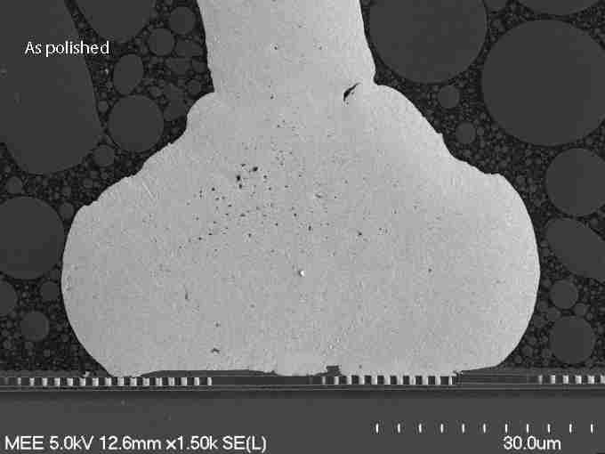
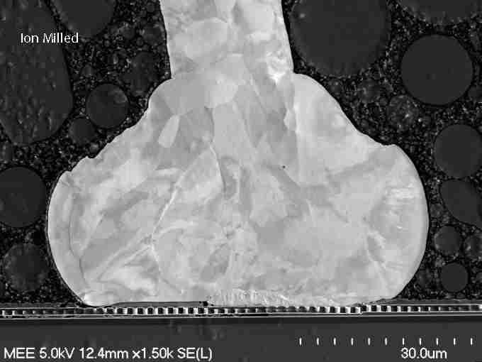
These images show a gold ball bond on an integrated circuit. In the top image we see the sample after it was mechanically prepared/polished. The next image was taken after the sample was ion milled.

- As polished

– As polished

 - Ion Milled

– Ion Milled

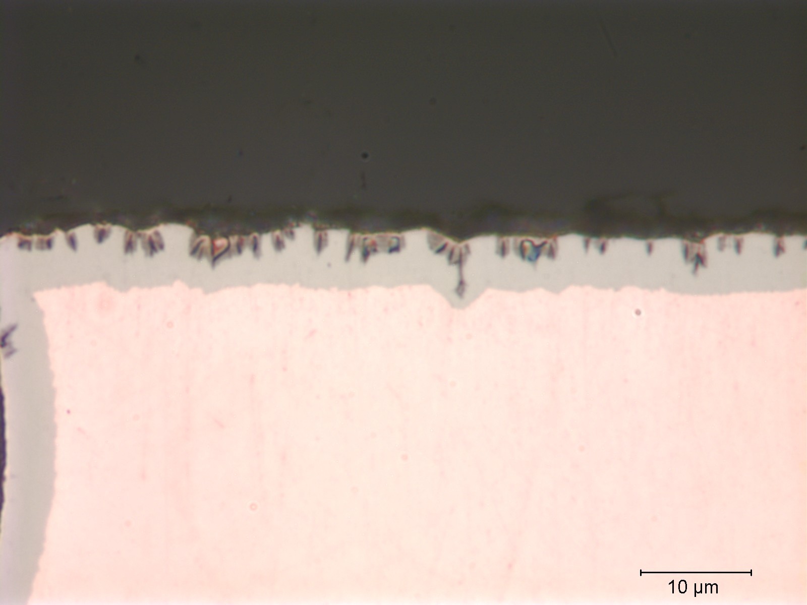
Microscopic inspection for device quality assurance, failure analysis, and materials characterization relies on optimum sample preparation to produce accurate and useful data. Good sample preparation for medical devices, semiconductors, microelectronics, and nano-materials has become more challenging in recent years due to high-technology materials, complex assemblies, and smaller components. Although mechanical cross sectioning, polishing, and chemical etching are sufficient for many applications, ion beam milling provides an additional level of quality and clarity for critical and difficult-to-prepare samples.